Pelvic Anatomy Vessels - Arteries Of The Pelvis Internal Iliac Pudendal Vesical Teachmeanatomy. All the pelvis viscera, i.e. There are four main arteries of the pelvis: The union of the internal and external iliac veins creates the common iliac vein, while the inferior epigastric vein drains into external iliac vein and anastomoses from the superior epigastric vein. The pelvic lymph nodes and vessels return lymph drained from pelvic organs, and follow a certain course that generally, but not reliably, follow the pattern of venous drainage of the pelvic structures. These veins of the pelvis correspond to the major pelvic arteries and share their names.
The two principal venous tributaries in the pelvis are the internal and external iliac veins, while the gastric veins dominate the abdomen. The femoral artery and femoral vein — two major blood vessels — travel through the pelvic bone. The obturator nerves and vessels are the lower limits of dissection of pelvic lymph nodes and lie between the external and internal iliac vessels. These structures are found on the posterolateral walls of the pelvic cavity. Superficial and caudal portions the middle gluteal, lateral rotators of the hip, and the adductor m.
Arteries And Lymphatics Of The Pelvis Dummies from www.dummies.com The obturator artery is the only artery arising laterally from the internal iliac artery. We think this is the most useful anatomy. The pelvis contains numerous structures, all supplied by the neurovascular structures of the pelvis, including nerves, arteries, veins, and lymph nodes. Hurd ww, bude ro, delancey jo, newman js. Here, we comprehensively analyzed intrapelvic vessel patterns. All the pelvis viscera, i.e. Anatomynote.com found pelvic wall blood vessels and nerves diagram from plenty of anatomical pictures on the internet. On each side of the pelvis, there are four routes for vessels to exit out of pelvis formed by the l5, s1, and s2 spinal nerve branches.
On each side of the pelvis, there are four routes for vessels to exit out of pelvis formed by the l5, s1, and s2 spinal nerve branches.
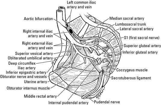
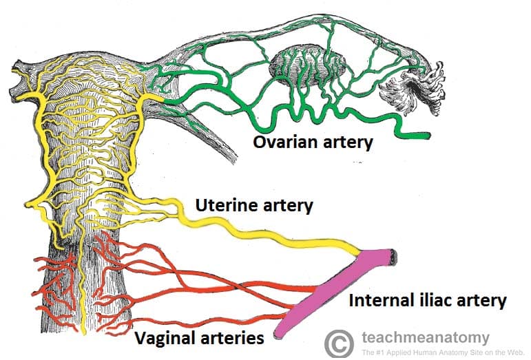
Main branches of the blood vessels and nerves of the uterus are located in this coat. In this dissection the veins, which follow the arteries closely, have been removed to simplify the picture. The femoral artery and femoral vein — two major blood vessels — travel through the pelvic bone. We hope this picture pelvic wall blood vessels and nerves diagram can help you study and research. The union of the internal and external iliac veins creates the common iliac vein, while the inferior epigastric vein drains into external iliac vein and anastomoses from the superior epigastric vein. The fibers of luschka (fl) are shown as they attach the paravaginal connective tissue to the sheath of the pubococcygeus. These travel down each leg, where they branch into internal and external iliac arteries. There are four main arteries of the pelvis: Knowledge of the vascular anatomy is essential to performing embolization and revascularization procedures in the male pelvis. Terminal parts of the digestive and urinary system; The external iliac vein, the internal iliac vein, and the common iliac vein. The internal iliac veins join the external iliacs to form the paired common iliac veins. Lymph nodes are usually clustered around the veins, and lymph vessels that come off these nodes ascend parallel to.
Lymphatic capillaries with fenestrated endothelial cell lining are blind ended tubes found in capillary bed. The internal iliac veins join the external iliacs to form the paired common iliac veins. Because the pelvis is in proximity to major blood vessels and organs, pelvic. The pelvic lymph nodes and vessels return lymph drained from pelvic organs, and follow a certain course that generally, but not reliably, follow the pattern of venous drainage of the pelvic structures. Terminal parts of the digestive and urinary system;
Arteries Of The Pelvis Internal Iliac Pudendal Vesical Teachmeanatomy from teachmeanatomy.info There are three major veins providing the venous drainage from the pelvic walls and viscera: On each side of the pelvis, there are four routes for vessels to exit out of pelvis formed by the l5, s1, and s2 spinal nerve branches. Anatomynote.com found pelvic wall blood vessels and nerves diagram from plenty of anatomical pictures on the internet. Pelvic spaces —the space between the bladder and the anterior portion of the pelvic walls is the perivesical space or space of retzius. These structures are found on the posterolateral walls of the pelvic cavity. The gonadal arteries arise directly from the aorta and carry blood to the gonads (testicles or ovaries). Normal anatomy the gonadal vessels refer to the testicular artery and testicular vein in males and the ovarian artery and ovarian vein in females. The internal iliac veins join the external iliacs to form the paired common iliac veins.
Larger of the two terminal branches of the internal iliac a.
Arteries and veins branch off from the femoral. In the pelvis, the abdominal aorta forks into two branches called common iliac arteries. Terminal parts of the digestive and urinary system; These vessels transport blood to and from each leg. This retrospective analysis included 81 patients that underwent colorectal surgery in our institution in 2016. We'll also remove the lining of peritoneum and pelvic fascia. For more anatomy content please follow us and visit our website: Normal anatomy the gonadal vessels refer to the testicular artery and testicular vein in males and the ovarian artery and ovarian vein in females. Main branches of the blood vessels and nerves of the uterus are located in this coat. Ramus s1 is separated from ramus s2 by the inferior gluteal vessels. The location of abdominal wall blood vessels in relationship to abdominal landmarks apparent at laparoscopy. The pelvis contains numerous structures, all supplied by the neurovascular structures of the pelvis, including nerves, arteries, veins, and lymph nodes. Larger of the two terminal branches of the internal iliac a.
There are four main arteries of the pelvis: Anatomynote.com found pelvic wall blood vessels and nerves diagram from plenty of anatomical pictures on the internet. The location of abdominal wall blood vessels in relationship to abdominal landmarks apparent at laparoscopy. The external iliac vein, the internal iliac vein, and the common iliac vein. This retrospective analysis included 81 patients that underwent colorectal surgery in our institution in 2016.
Functional Anatomy Of Pelvic Organs Springerlink from media.springernature.com Drapes over the fallopian tubes, uterus, ovaries, and blood vessels extends from the lateral walls of the uterus to the sidewalls of the pelvis provides a small amount of support for the uterus creates the retrouterine and vesicouterine pouches Pelvic spaces —the space between the bladder and the anterior portion of the pelvic walls is the perivesical space or space of retzius. The iliac and femoral vessels run anterior to the bones of the pelvis, making them most apt to injury in trauma to the tissue or bones of the anterior pelvis. We hope this picture pelvic wall blood vessels and nerves diagram can help you study and research. Here, we comprehensively analyzed intrapelvic vessel patterns. The external iliac vein, the internal iliac vein, and the common iliac vein. Lymph nodes are usually clustered around the veins, and lymph vessels that come off these nodes ascend parallel to. The two principal venous tributaries in the pelvis are the internal and external iliac veins, while the gastric veins dominate the abdomen.
Its arterial supply is largely via the internal iliac artery, with some smaller arteries providing additional supply.
The iliac and femoral vessels run anterior to the bones of the pelvis, making them most apt to injury in trauma to the tissue or bones of the anterior pelvis. The pelvis is the sturdy ring of bones located at the base of the spine. On each side of the pelvis, there are four routes for vessels to exit out of pelvis formed by the l5, s1, and s2 spinal nerve branches. This retrospective analysis included 81 patients that underwent colorectal surgery in our institution in 2016. The two principal venous tributaries in the pelvis are the internal and external iliac veins, while the gastric veins dominate the abdomen. The internal iliac veins join the external iliacs to form the paired common iliac veins. Anatomynote.com found pelvic wall blood vessels and nerves diagram from plenty of anatomical pictures on the internet. Atlas of pelvic anatomy and gynecologic surgery, 3rd ed, baggish ms, karram mm (eds), elsevier saunders, st. In this dissection the veins, which follow the arteries closely, have been removed to simplify the picture. These veins of the pelvis correspond to the major pelvic arteries and share their names. Drapes over the fallopian tubes, uterus, ovaries, and blood vessels extends from the lateral walls of the uterus to the sidewalls of the pelvis provides a small amount of support for the uterus creates the retrouterine and vesicouterine pouches These structures are found on the posterolateral walls of the pelvic cavity. Hurd ww, bude ro, delancey jo, newman js.
Its arterial supply is largely via the internal iliac artery, with some smaller arteries providing additional supply pelvic anatomy. Because the pelvis is in proximity to major blood vessels and organs, pelvic.
0 Comments:
Posting Komentar