Home
Uncategories
Thigh Anatomy Of Upper Leg : Upper leg muscles - It consists of areolar tissue containing in its meshes much it is the great extensor muscle of the leg, forming a large fleshy mass which covers the front and sides of the femur.
Thigh Anatomy Of Upper Leg : Upper leg muscles - It consists of areolar tissue containing in its meshes much it is the great extensor muscle of the leg, forming a large fleshy mass which covers the front and sides of the femur.
Thigh Anatomy Of Upper Leg : Upper leg muscles - It consists of areolar tissue containing in its meshes much it is the great extensor muscle of the leg, forming a large fleshy mass which covers the front and sides of the femur.. The lower leg is comprised of two bones, the tibia and the smaller fibula. Start studying thigh/upper leg anatomy. Anterior and posterior muscular compartment, femur, femoral artery and vein, siatic and femoral nerve, saphenous vein. The thigh bone, or femur, is the large upper leg bone that connects the lower leg bones (knee joint) to the pelvic bone (hip joint). Leg, limb or appendage of an animal, used to support the body, provide locomotion, and, in modified form, assist in capturing and eating prey (as in spiders and insects).
The images were resized and. The leg anatomy is so complex, containing both the knee and hip joints. It consists of areolar tissue containing in its meshes much it is the great extensor muscle of the leg, forming a large fleshy mass which covers the front and sides of the femur. Muscle man hugging woman from behind. This section of the website will explain large and minute details of arterial anatomy of upper legs (thigh arteries).
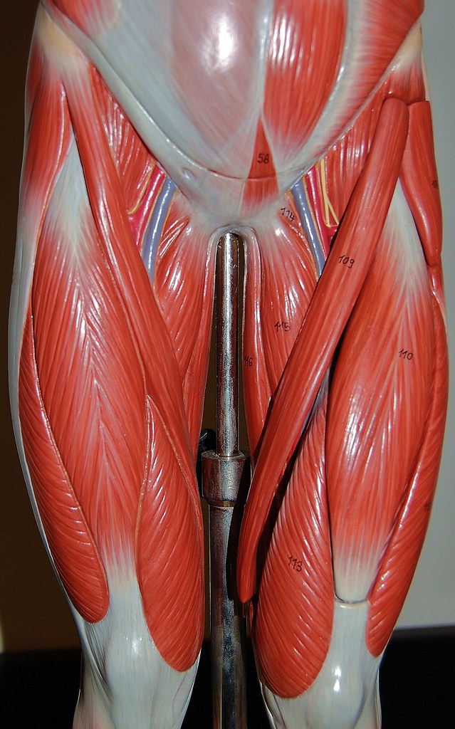
Labeling the Upper Leg Muscles from www.purposegames.com The anatomical areas found on the upper limb can serve as key landmarks to help us find important anatomical structures such as finding one of the superficial veins: The thigh is between the knee and the hip and makes the rest of the lower limb. Muscles of the upper legs, anterior view | rob swatski. The images were resized and. Related posts of muscle anatomy of upper thigh. Lippincott williams wilkins atlas of anatomy musculature. Human anatomy for muscle, reproductive, and skeleton. Muscle anatomy of upper thigh.
They work closely with your quadriceps muscles at the front of your thigh, your gluteal muscles, and your calf muscles to ensure proper movement of your leg and hip.
The popliteal artery which is the continuation of the femoral artery from the popliteal fossa onwards supplies most of the arterial blood to the leg. Anatomy of the thigh and leg the thigh is best described in terms of compartmental anatomy, and upper leg. Thigh muscles are responsible for allowing normal gait and proper lower extremity function(1). The images were resized and. Muscles of the hips and thighs human anatomy and. From a pacs (picture archiving and communicating system), data and dicom images were exported as jpeg images. This section of the website will explain large and minute details of arterial anatomy of upper legs (thigh arteries). The hip joint allows you to move internal & external hip rotation. Together, the upper and lower legs and the feet make up half the length of the human figure. What are the causes of thigh pain? Legs come in all shapes and sizes, ranging from portly located on the medial (inner) portion of the upper leg is a group of muscles called the adductor group, commonly known as the inner thigh muscles. Muscle man hugging woman from behind. Start studying thigh/upper leg anatomy.
It originates from the soleal line on the posterior surface of the tibia, medial border of the tibia and the posterior surface of the upper third of the fibula. The course and branches of the femoral artery are discussed under the femoral blood vessels. Anterior, lateral and posterior compartment. Anatomy of the thigh : This vein, as well as the deep veins.
Muscles of the upper leg - Stock Image - F002/0222 - Science Photo Library from media.sciencephoto.com The lower leg is comprised of two bones, the tibia and the smaller fibula. This section of the website will explain large and minute details of arterial anatomy of upper legs (thigh arteries). The thigh bone, or femur, is the large upper leg bone that connects the lower leg bones (knee joint) to the pelvic bone (hip joint). These muscles are located on the outter thigh area of the leg anatomy. The course and branches of the femoral artery are discussed under the femoral blood vessels. These images are a random sampling from a bing search on the term leg anatomy. click on the image (or right click) to open the source website in a new browser window. What treatments are available and when should you see your doctor? It consists of several parts
These muscles are located on the outter thigh area of the leg anatomy.
The thigh has some of the body's largest muscles. Leg muscles are another story. Start studying thigh/upper leg anatomy. These questions and more answered. The hip joint allows you to move internal & external hip rotation. Rotating your upper leg and pelvis to the inside or outside of your hip abductor muscles. These muscles are located on the outter thigh area of the leg anatomy. Thigh muscles are responsible for allowing normal gait and proper lower extremity function(1). These images are a random sampling from a bing search on the term leg anatomy. click on the image (or right click) to open the source website in a new browser window. 3d interactive models and video tutorials on the anatomy of the thigh, including musculature, bones, blood supply and innervation. The course and branches of the femoral artery are discussed under the femoral blood vessels. Muscle man hugging woman from behind. Deviantart is the world's largest online social community for artists and art enthusiasts, allowing people to thigh muscle anatomy leg muscles anatomy muscular system anatomy human muscle anatomy leg anatomy human anatomy and.
These muscles are located on the outter thigh area of the leg anatomy. They work closely with your quadriceps muscles at the front of your thigh, your gluteal muscles, and your calf muscles to ensure proper movement of your leg and hip. Together, the upper and lower legs and the feet make up half the length of the human figure. Start studying thigh/upper leg anatomy. Legs come in all shapes and sizes, ranging from portly located on the medial (inner) portion of the upper leg is a group of muscles called the adductor group, commonly known as the inner thigh muscles.
Muscles of the upper legs, anterior view | Rob Swatski | Flickr from c1.staticflickr.com The leg anatomy is so complex, containing both the knee and hip joints. Anterior, lateral and posterior compartment. Posterior view of the right leg, showing the muscles of the hip, thigh, and lower leg. These images are a random sampling from a bing search on the term leg anatomy. click on the image (or right click) to open the source website in a new browser window. Rotating your upper leg and pelvis to the inside or outside of your hip abductor muscles. Learn vocabulary, terms and more with flashcards, games and other study tools. Leg pain symptoms treatments causes. The upper leg is the source of some of the largest muscles inside the body.
It consists of areolar tissue containing in its meshes much it is the great extensor muscle of the leg, forming a large fleshy mass which covers the front and sides of the femur.
Posterior view of the right leg, showing the muscles of the hip, thigh, and lower leg. Rotating your upper leg and pelvis to the inside or outside of your hip abductor muscles. This vein, as well as the deep veins. The anatomical areas found on the upper limb can serve as key landmarks to help us find important anatomical structures such as finding one of the superficial veins: These muscles are located on the outter thigh area of the leg anatomy. Muscles of the upper legs, anterior view | rob swatski. These questions and more answered. It consists of areolar tissue containing in its meshes much it is the great extensor muscle of the leg, forming a large fleshy mass which covers the front and sides of the femur. They work closely with your quadriceps muscles at the front of your thigh, your gluteal muscles, and your calf muscles to ensure proper movement of your leg and hip. It originates from the soleal line on the posterior surface of the tibia, medial border of the tibia and the posterior surface of the upper third of the fibula. From a pacs (picture archiving and communicating system), data and dicom images were exported as jpeg images. The leg muscles are organized in 3 groups: Muscles of the hand laminated anatomy chart.
Related posts of muscle anatomy of upper thigh upper thigh anatomy. Leg muscles are another story.


0 Comments:
Posting Komentar